
|
Institute for Biology and Medical Genetics, First Faculty of Medicine, Charles University, General Teaching Hospital, Prague |

|
Biology and Genetics - practical exercises
Week 6 - Mitosis
Task 1
Your first task is to study the microphotograph of the mitosis. Note the size and morphological differences of the chromosomes. In order to vizualize the chromosomes - various staining - banding methods are used (in this case G-banding method).
|
Left-click to show chromosome:
Chromosome 1
Chromosome 2 Chromosome 3
Chromosome 4
Chromosome. 5
Chromosome 6
Chromosome 7 Chromosome 8 Chromosome 9 Chromosome 10 Chromosome 11 Chromosome 12
Chromosome 13
Chromosome 14 Chromosome 15
Chromosome 16
Chromosome 17 Chromosome 18
Chromosome 19
Chromosome 20
Chromosome 21
Chromosome 22
Gonosomes
Clear all |
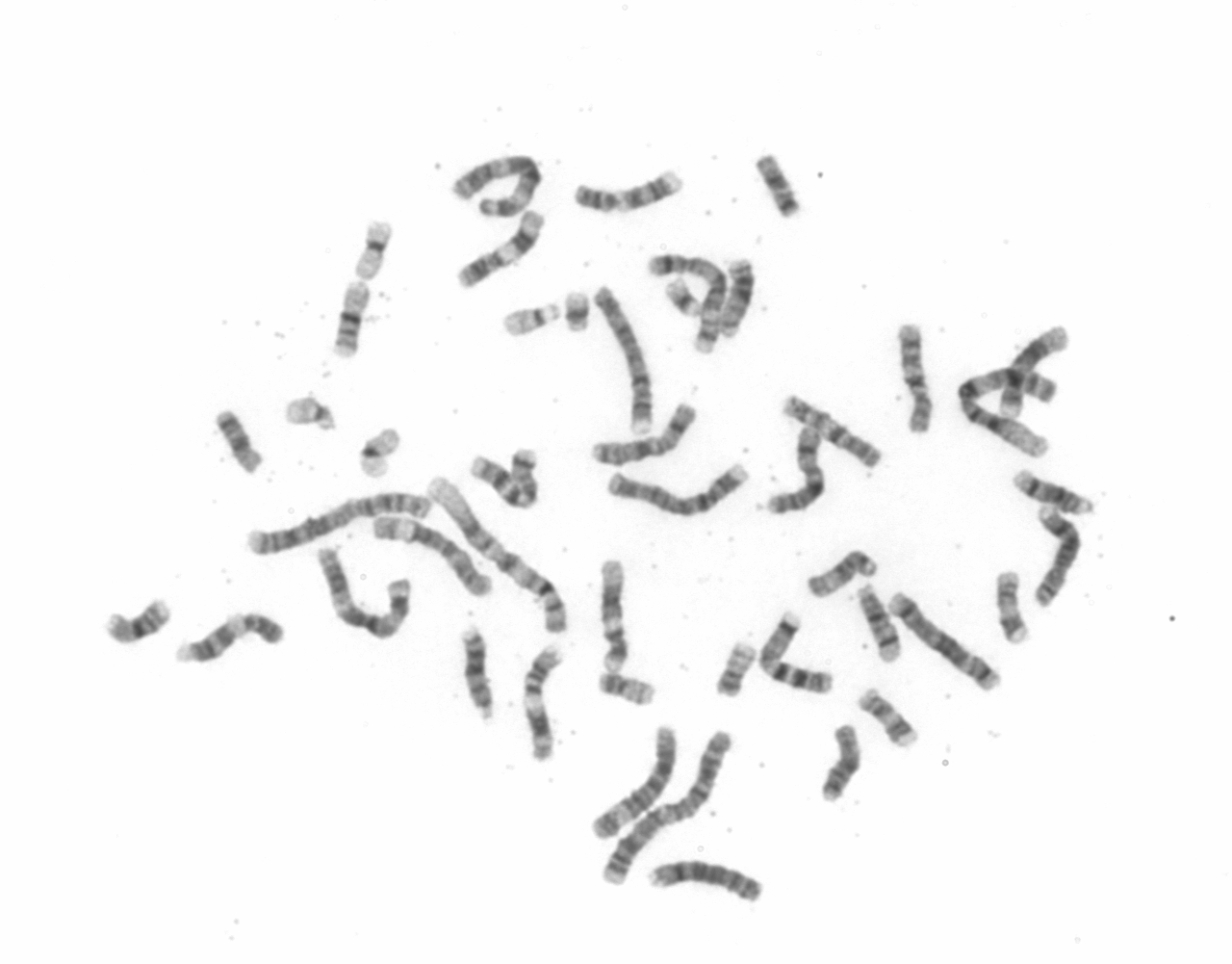 |
Task 2
Your next task is to :
- count all chromosomes within the mitosis (metaphase)
- determine the gender of the karytyped person.
If you get 4 small chromosomes - then the chromosome Y is not present - so the karyotype is female.
But if you get 5 small chromosomes (including the Y chromosome), then the karyotype is male.
For better orientation you can see the ideograms of all human chromosomes - .
The solution will be shown in the presentation.
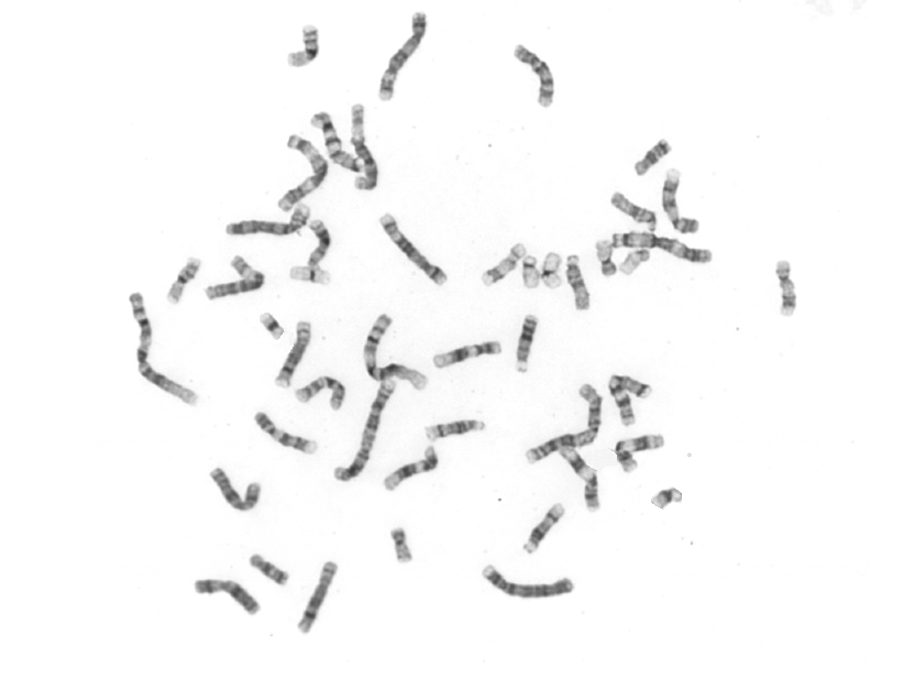
Task 3
Again, you have to count all chromosomes and determine the gender of the karyotyped person.
The solution will be shown in the presentation.
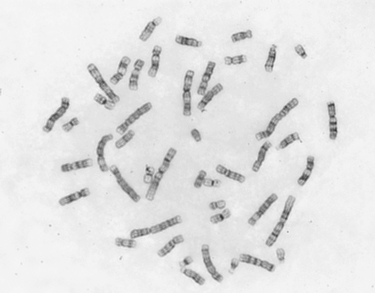
Task 4
Your next task is to make a karyotype - in the same way like the clinical cytogenetics in the practice. You have a little bit easier situation, because one of each chromosome pairs has already been placed. Your task is to find the second chromosome for each chromosome pair and complete the work.
Make a karyotype - Flash animation (University of Utah).